|
")
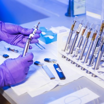
A femtosecond laser is a laser which emits optical pulses with an extremely short duration, known as of femtoseconds (1 femtosecond equals 10 in the -15 seconds). The femtosecond laser used for this treatment produces brief, high energy light pulses that cut through the target cell. The laser heat does not spread around to harm nearby healthy cells. There is hope in the medical community that this device could be used for accurate demolition of various types of unhealthy tissue. This would include small tumors of the vocal cords, cancer cells left behind after the removal of solid tumors, individual cancer cells scattered throughout the brain, or other tissue and plaque in arteries.
Femtosecond lasers have been developed by several companies around the world but until recently have been heavy and cumbersome. Ben-Yakar’s laboratory has created a microscope system that is capable of delivering femtosecond laser pulses up to 250 microns deep inside a tissue. This system has a tiny flexible probe that can focus light pulses to a spot smaller than a human cell.
Ben-Yakar hopes that in a few years the probe’s 15-millimeter diameter will shrink three fold, thus making it match endoscopes that are engaged in laparoscopic surgeries today. She also considers making the probe tip disposable so it could be used in operations on people with have infectious diseases or destroying deadly viruses and other biomaterials.
|
")
During the development of the miniature-surgery system, Ben-Yakar worked in collaboration with Olav Solgaard at Stanford University’s electrical engineering department in order to incorporate a miniaturized scanning mirror. Another addition to the system was made by Ben-Yakar and her graduate student Chris Hoy; they used a novel optic-fiber cable that can withstand the light pulses the laser sends out. While trying to make the laser intensity easier to manage, they stretched the light pulses into longer, weaker pulses that travel through the fiber. Then, the fiber’s properties were used for reconstructing the light into more intense, short light pulses before they entered the tissue.
In this study, Ben-Yakar targeted breast cancer cells in three-dimensional bio-structures that mimicked the optical properties of breast tissue. The laser has also been tested on laboratory-grown layered cell structures that mimic different tissues.
An additional line of research Ben-Yakar is currently engaging is the use of nanoparticles to focus the light energy on targeted cells. She recently demonstrated that gold nanoparticles can function as magnifying lenses, thus increasing the amount of laser light reaching the cells by at least an order of magnitude. When this technique is fully developed it would be possible to target hundreds of effected cells in a single tissue at a time without affecting the healthy cells.
TFOT has recently covered several other stories on new treatments for cancer. One such story tells of a new infrared camera developed by researchers from the Argonne National Laboratory which can take images of the head and neck and predict which patients are likely to develop severe side effects due to anti-cancer treatments and adapt the treatment accordingly. Another story tells of a personalized melanoma vaccination developed by Hadassah Medical Organization and the Hebrew University in Jerusalem.
More information on the new laser can be found on the Texas University website.