|
")
Metastatic cells make a long journey in order to reach the blood flow and spread to distant organs. In order to be able to make this journey, the cells need to be extremely flexible. Becoming soft and flexible is not an easy task and requires the cancer cells to undergo additional mutations. This research provides proof that this property makes the metastatic cells easily distinguishable from normal cells. This procedure is essential when there’s a case of fluids buildup in the chest and abdominal cavities, which sometimes occurs due to the presence of metastatic cells.
Using the available methods today, doctors can’t easily distinguish normal cells and metastatic cells. Potentially, if physicians could easily know which of the cells are cancerous, they could detect metastasis at an earlier stage, lowering patients’ death rates. Today, in a case of fluid buildup, doctors examine the cells in the fluids using a light microscope and by dying the cells with certain chemicals. These methods are old and usually are not accurate enough to distinguish a cancer cell from a normal cell. The UCLA research has succeeded in finding a differing character, the cell’s softness, which will help doctors recognize a cancer cell when it’s present in a sample.
The softness of the cells is measured using an AFM (atomic force microscope), and the research has proven that this measurement can determine with a high degree of confidence which cells are metastatic and which are normal. The atomic force microscope is a microscope that can “sense” the surface of the specimen by reading changes in the forces applied to it. The resolution of the microscope is more than thousand times higher than that of a conventional optical microscope, allowing it to distinguish objects measuring fractions of a nanometer (one billionth of a meter), and even to tell apart individual atoms. In the UCLA research project, the microscope is used as a gentle probe that pressures the surface of the cell. The resistance of the cell’s surface is measured and displayed as output. In experiments conducted on samples of liquids taken from patient’s cavities the measurement was found to be highly useful in detecting metastatic cells.
The results of the experiments showed that the softness of the cell’s surface can be used to tell a metastatic cell from a normal cell. The experiment showed that this attribute is highly distinctive and that this scan is applicable and can be used as common practice in analyzing liquids buildups in cavities.
TFOT has recently covered several other devices that can help detect cancer outside the body by using different characteristics of cancer cells. These include the T-rays detector developed by the U.S. Department of Energy’s Argonne National Laboratory, a ‘lab on a chip’ to detect oral cancer developed at the Texas University, and the nanocytometer chip developed at the University of California, Berkeley for locating cancer cells in the blood stream.
More information is available on the UCLA website.
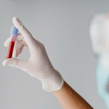
Top image: An artist’s depiction of an atomic force microscope probe (Credit: Arizona State University).