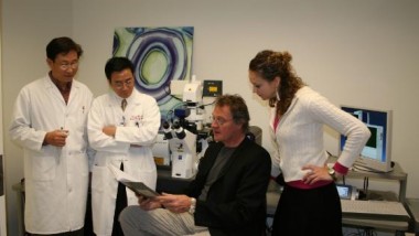
Department Head, Department of Biomedical Engineering and Project Lead – Rensselaer Polytechnic Institute Shoulder x-ray. Rensselaer Polytechnic Institute has developed a technique using “laser-capture microscopy” that allows researchers to collect large amounts of biochemical information from nanoscale bone samples. Since ...
When Softness isn’t a Virtue
A team of researchers from the University of California in Los Angeles (UCLA) has developed a method to distinguish between metastatic cancer cells and normal cells. The method utilizes an atomic force microscope (AFM) to measure the softness of a ...
ISAM – Computed Image Revolution
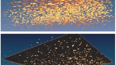
Researchers at the University of Illinois at Urbana-Champaign have developed a novel computational image-forming technique for optical microscopy, called ISAM. The new technique is capable of producing three-dimensional images even from blurry, out-of-focus data. The new tool will improve the ...
MEMS-based Confocal Microscopy
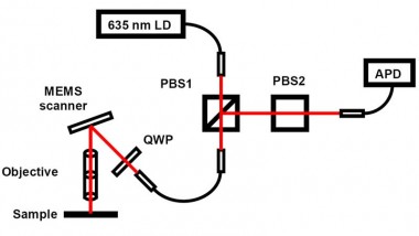
A new MEMS based scanning technology offers a different possibility for conducting confocal microscopy imaging in confined sections of a living human body. Professor Rebecca Kortum Confocal microscopy is an imaging technique that offers high-resolution imaging of living tissues (in ...


