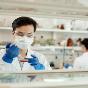
High-resolution imaging’s a cornerstone of life sciences, letting researchers peek into cells like never before. It’s all about watching living systems in real time, decoding cellular structures with jaw-dropping clarity. New microscopy tools are shaking things up in labs, and the future’s looking bright. This tech’s evolving fast, and it’s kind of a big deal for pushing science forward.
Spinning Disc Microscopy: The Go-To Star
One standout in this space is spinning disc confocal microscopy. It snaps crystal-clear images of live cells, and researchers can’t get enough of it. Compared to older methods, it’s gentler and way faster. It’s less stressful on samples, which is huge for keeping cells happy during experiments. Labs lean on it to track proteins or cell functions, and it’s a total game-changer for studying dynamic processes.
Live-Cell Imaging That Doesn’t Wreck Samples
Old-school imaging tools could be rough on cells. Too much light exposure stressed samples, sometimes even killing them. That made long-term studies tricky, and the data wasn’t always reliable. Spinning disc microscopy flips the script by dialing down light intensity. Researchers can snap images over and over without harming cells, catching live processes as they unfold. It’s like getting a front-row seat to biology in action, with sharper insights into how cells tick.
Software That’s Smarter Than Ever
Fancy hardware’s only half the story—software’s where the magic really happens. Modern imaging systems come with built-in analysis tools that handle massive piles of image data. They streamline workflows, which is a lifesaver in busy labs. These programs can spot tiny shifts in cell behavior, saving researchers from squinting at screens for hours.
Some systems even use machine learning to up their game. They catch patterns humans might miss, like subtle protein movements. Tools for image stitching let you piece together big, detailed views or build 3D models from flat images. It’s pretty darn cool, making analysis faster and way more precise.
Multiplex Imaging: More Bang for Your Buck
Researchers are always hungry for more data from each sample, and multiplex imaging delivers. It lets you tag different targets—like proteins or structures—with unique fluorescent markers, so you can watch multiple things at once. No need to run the same experiment over and over. You can compare variables in one go, which saves time and gives richer results.
This trick’s a favorite in cancer research and drug development. It shows how cells react to treatments, revealing connections that’d be tough to spot otherwise. It’s efficient, cost-effective, and a no-brainer for labs wanting to stretch their samples further.
Compact Systems for Any Lab
High-end microscopes used to demand big spaces and bigger budgets. Not anymore. Compact systems are hitting the scene, packing high-res power into portable packages. They’re perfect for smaller labs or even fieldwork, where space is tight.
These setups are user-friendly, with plug-and-play designs and touchscreen controls. Many sync with cloud platforms, so you can store and share data remotely. That makes collaborating a breeze—upload images, and your team across the globe can dive in. It’s a practical way to scale up without a massive investment.
Cryo-Imaging: Freezing for Precision
Cryogenic imaging’s making waves, especially in structural biology. It flash-freezes samples to lock in their natural shape, ideal for studying delicate proteins or tiny molecules. Cryo-electron microscopy’s a big player here, offering insane detail.
Newer tools mix cryo-imaging with fluorescent tags, blending structural and functional views. You can see where molecules are and what they’re doing—like mapping a virus or enzyme in action. These systems aren’t cheap, but their accuracy’s unmatched, making them worth it for cutting-edge work.
Tying It All Together with Data
Imaging’s most powerful when it plays nice with other methods. Labs are pairing microscopy with genomics or proteomics for a fuller picture of biology. You’re not just seeing structures—you’re linking them to what they do, which is huge for discoveries.
AI’s a key teammate here. It crunches massive image sets, spots trends, and points researchers to what matters. Machine learning can even suggest where to focus next, cutting down on grunt work. This combo’s becoming standard, letting labs move faster and with more confidence.
What’s Next?
High-resolution imaging’s shaping where life sciences are headed. Tools like spinning disc confocal microscopy are leading the charge, letting researchers see more without trashing samples. Smarter software’s boosting analysis, while compact systems make this tech accessible to more labs. Cryo-imaging’s pushing the limits of detail, and tying it all to AI and other data streams is unlocking new insights.
The future’s packed with potential. We’ll see even smaller, cheaper systems with more automation—think AI tweaking images on the fly. Labs that jump on these tools now will stay ahead, cranking out discoveries that matter. It’s an exciting time, and high-res imaging’s at the heart of it, helping scientists unravel biology’s biggest mysteries.