|
")
Optical traps are devices that focus light on individual particles. While optical traps have been around for years, they traditionally require large groups of lasers and microscopes to focus that light. Dr. Kenneth Crozier and his colleagues at Harvard have designed three different simple optical traps that fit on standard silicon chips, a massive miniaturization. These chips interface with existing microfluids developed to diagnose cancer and sort the different cells present within a fluid, gently attracting individual cells to the surface of the optical trap chips for evaluation.
Developers build two of the three designs currently under evaluation upon standard silicon chips. One uses a ring pattern five micrometers in diameter and the other a series of bullseye patterns. In both cases, photons generated when laser light illuminates the patterns draw in particles from a flowing liquid above the chip. In addition, the laser focuses the light in a manner similar to that of a microscope and can possibly generate a 3-dimensional image of the captured cell. Scientists could then use these images to identify cancer cells and other diseases.
The third design uses tapered gold films placed on silicon chips. When illuminated with light passed through a small prism, the gold strips generate surface waves called plasmons. The plasmons can pull down particles and push them along the gold surface. The speed of these particles can be controlled by adjusting the angle of the light applied. This structure should be particularly well suited for cell sorting, a task now currently performed with bulky flow cytometers.
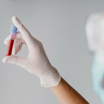
TFOT has previously reported on a variety of other potential diagnostic tools for cancer, including a new contrast agent designed to help improve tumor detection in MRI scans, a new way to scan blood to find cancer in its very earlier stages, infrared sensors that can detect cancer markers in the breath of patients, and radio sensors that can help detect esophageal cancer.
Read more about Crozier’s research at his Harvard University group page.