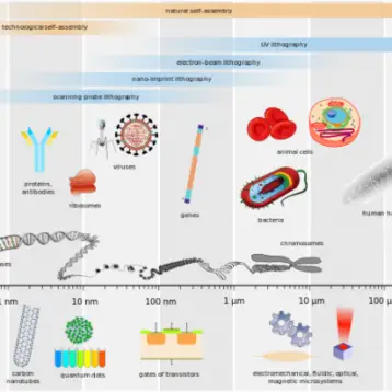
The new device is tiny – even smaller than some viruses – and in comparison to contemporary probes, it is 100-times smaller. Thanks to its nanotechnology-based design, the device allows scientists to probe cells without damaging them. Moreover, the reduction in size did not cause any accuracy reduction, and therefore the data reliability is maintained.
Professor Charles M. Lieber from Harvard, the study’s author, said: “Our use of these nanoscale field-effect transistors, or nanoFETs, represents the first totally new approach to intracellular studies in decades, as well as the first measurement of the inside of a cell with a semiconductor device. The nanoFETs are the first new electrical measurement tool for intracellular studies since the 1960s, during which time electronics have advanced considerably.”
According to the recent paper, appearing in the journal Science, nanoFETs could be used to measure ion flux or electrical signals in cells, particularly neurons. The device could also be fitted with receptors or ligands to probe for the presence of individual biochemicals within a cell.
The size of human cells varies; it ranges from about 10 microns (millionths of a meter) for nerve cells to 50 microns for cardiac cells. While current probes measure up to 5 microns in diameter, nanoFETs are several orders of magnitude smaller: less than 50 nanometers (billionths of a meter) in total size, with the nanowire probe itself measuring just 15 nanometers in diameter.
The new probe provides two more benefits aside from its minimal size. One is easy insertion of nanoFETs into cells, possible by coating the structures with a phospholipid bilayer — the same material cell membranes are made of — the devices are easily pulled into a cell via membrane fusion, a process related to that used to engulf viruses and bacteria. “This eliminates the need to push the nanoFETs into a cell, since they are essentially fused with the cell membrane by the cell’s own machinery,” said Lieber.
The second benefit is the introduction of triangular “stereocenters” — essentially, fixed 120-degree joints — into nanowires, structures that had previously been rigidly linear. These stereocenters, analogous to the chemical hubs found in many complex organic molecules, introduce kinks into 1-D nanostructures, transforming them into more complex forms. According to Lieber, it means that insertion of nanoFETs is not nearly as traumatic to the cell as current electrical probes. “We found that nanoFETs can be inserted and removed from a cell multiple times without any discernible damage to the cell,” he said. “We can even use them to measure continuously as the device enters and exits the cell.”
This innovative study was co-authored; Professor Lieber’s team members are Bozhi Tian, Tzahi Cohen-Karni, Quan Qing, Xiaojie Duan, and Ping Xie, all of Harvard’s Department of Chemistry and Chemical Biology and School of Engineering and Applied Sciences. Furthermore, the work was sponsored by the National Institutes of Health and the McKnight Endowment Fund for Neuroscience.
TFOT also covered the single-atom transistor, built out of a single phosphorus atom in silicon, and the nano targeted drug delivery, studied at the California NanoSystems Institute and Northwestern University.
For more information about the nanoscale transistors that probe cells, see Harvard’s official press release.


