|

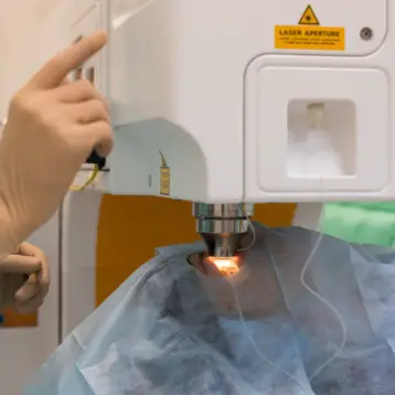
Confocal microscopy is an imaging technique that offers high-resolution imaging of living tissues (in vivo). However, in order to generate the image, an optical beam must be focused and scanned across the sample at high speed. Professor Rebecca Richards Kortum of Stanford University and her team, proposed a microelectromechanical (MEMS) method to conduct the scanning using mirrors.
Mirrors with sub-millimeter dimensions can be batch fabricated from a silicon-on-insulator (SOI) wafer, following multiple etching steps to leave a tilting mirror surface attached to the fixed substrate. Such mirrors with rotating capability will enable scanning of one point while a different mirror orientation will collect light from a different area. A single gimbal-mounted surface is capable of rotating round orthogonal axes and can scan an optical beam over a two-dimensional raster pattern, eliminating the need to cascade successive one dimensional scanners.
The main promise of this new technology is the conducting of confocal imaging during routine clinical use. In order for that to happen, further improvements like minimizing the motion included in the imaging process, and improving the field-of-view and resolution (The axial and lateral resolution values for the system were experimentally measured to be 9.55 micrometer and 0.83 micrometer respectively, with a 140 micrometer x 70 micrometer field of view) will need to be made.
|

The team’s objectives in the project are to design, construct, and test a fiber optic endoscope to obtain confocal reflected light images of living human epithelial tissues. Epithelium cells are one of four primary body tissues which can be found in the skin, mucous membranes lining the inside of mouths, the lungs, the gastrointestinal tract, and the reproductive and urinary tracts, as well as in other organs.
The team also aims to develop techniques to interpret the resulting tissue images and yield diagnostic information based on sub-cellular morphologic and biochemical changes. The application of the resulting system will be to image epithelial tissues to aid medical staff in diagnosis, and potentially to enable screening of epithelial pre-cancers such as oral, uterine, urinary bladder, colon and other types of cancers.
More information on the Stanford research can be found here.